OBJECTIVES:
To reveal bacteria that stay in yogurt such as Streptococcus Thermophilus and Lactobacillus Bulgaricus and to know how to do the Gram Stain in order to reveal them.
MATERIAL
- Crystal violet
- Ethanol
- Distilled water
- Safranin
- Three beakers
- Dropper
- Bunsen burner
- Lighter
- Forceps
- Slide and coverslide
- Dissection needle
- Iodine (Lugol)
- Petri dish
PROCEDURES
Take the dissection needle and burn it in the Bunsen burner and then spread it on the yogurt.
Put the yogurt on a slide, then take a dropper and throw a few drops of Chrystal violet, wait for 1 minute and 30 seconds. After that clean the slide with some distilled water.
Now do the same with Iodine, throw on it few drops and wait for 1 minute, then clean it with water again.
Wash it well with ethanol.
Now you have to dye the slide with safranin and to put again some water, after that add the coverslide and put it into the microscope.
RESULTS

QUESTIONS
EXPLANATION OF GRAM STAIN
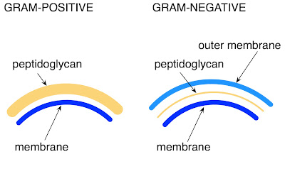
Gram-positive bacteria have a thicker peptidoglycan (disaccharides and amino acids) cell wall than gram-negative bacteria. The latter contain a layer of lipopolysaccharide (lipids and polysaccharides) as part of their cell wall. When applied to both gram-positive and gram-negative cells, crystal violet and then iodine readily enter the cells. Inside the cells, the crystal violet and iodine combine to form the crystal violet-iodine (CV- I) complex. This complex is larger than the crystal violet molecule that entered the cells and because of its size, it cannot be washed out of the intact peptidoglycan layer of gram-positive cells by alcohol. Consequently, gram-positive cells retain the color of the crystal violet dye. In gram-negative cells, however, the alcohol wash disrupts the outer lipopolysaccharide layer and the CV- I complex is washed out through the thin layer of peptidoglycan . As a result, gram negative cells are colorless until counterstained with safranin, after which they are pink.

OBSERVATION OF G+ OR G-
The first membrane belongs to Gram + and the second to Gram -.
No hay comentarios:
Publicar un comentario